
Home
|
Products
|
9789356962026
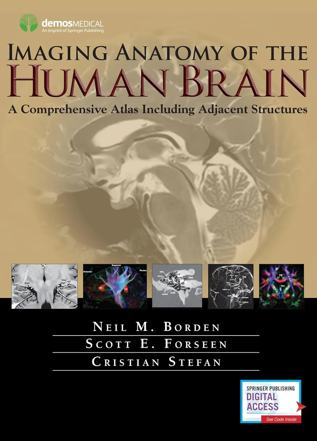
Imaging Anatomy of the Human Brain | Hardcover
by Borden
Highlights

9781936287741
ISBN
Borden
Author

464
Pages

2000 gm
Weight

English
Language

2016
Year

1st Edition
Edition

Hardcover
Binding
₹14252
₹15836
The most precise, cutting-edge images of normal cerebral anatomy available today are the centerpiece of this spectacular atlas for clinicians, trainees, and students in the neurologically-based medical and non-medical specialties. Truly an atlas for the 21st century, this comprehensive visual reference presents a detailed overview of cerebral anatomy acquired through the use of multiple imaging modalities including advanced techniques that allow visualization of structures not possible with conventional MRI or CT. Beautiful colour illustrations using 3-D modeling techniques based upon 3D MR volume data sets further enhances understanding of cerebral anatomy and spatial relationships. The anatomy in these colour illustrations mirror the black and white anatomic MR images presented in this atlas.Written by two neuroradiologists and an anatomist who are also prominent educators, along with more than a dozen contributors, the atlas begins with a brief introduction to the development, organization, and function of the human brain. What follows is more than 1,000 meticulously presented and labelled images acquired with the full complement of standard and advanced modalities currently used to visualize the human brain and adjacent structures•including MRI, CT, diffusion tensor imaging (DTI) with tractography, functional MRI, CTA, CTV, MRA, MRV, conventional 2-D catheter angiography, 3-D rotational catheter angiography, MR spectroscopy, and ultrasound of the neonatal brain. The vast array of data that these modes of imaging provide offers a wider window into the brain and allows the reader a unique way to integrate the complex anatomy presented. Ultimately the improved understanding you can acquire using this atlas can enhance clinical understanding and have a positive impact on patient care. Additionally, various anatomic structures can be viewed from modality to modality and from multiple planes.This state-of-the-art atlas provides a single source reference, which allows the interested reader ease of use, cross-referencing, and the ability to visualize high-resolution images with detailed labeling. It will serve as an authoritative learning tool in the classroom, and as an invaluable practical resource at the workstation or in the office or clinic.Key Features:Provides detailed views of anatomic structures within and around the human brain utilizing over 1,000 high quality images across a broad range of imaging modalitiesContains extensively labeled images of all regions of the brain and adjacent areas that can be compared and contrasted across modalitiesIncludes specially created colour illustrations using computer 3-D modeling techniques to aid in identifying structures and understanding relationshipsGoes beyond a typical brain atlas with detailed imaging of skull base, calvaria, facial skeleton, temporal bones, paranasal sinuses, and orbitsServes as an authoritative learning tool for students and trainees and practical reference for clinicians in multiple specialties
Online store of medical books
Discover a comprehensive range of medical books at our online store. From anatomy and physiology to the latest clinical guidelines, we've got you covered.
Trusted by students, educators, and healthcare professionals worldwide. Browse top publishers and expert-authored titles in every medical specialty. Enjoy fast shipping, secure payments, and easy returns. Your one-stop destination for quality medical knowledge at your fingertips.
Whether you're preparing for exams or expanding your clinical expertise, our curated collection ensures you have the right resources at hand. Dive into detailed illustrations, case studies, and up-to-date research that enhance your understanding and practical skills.
We regularly update our inventory to include the latest editions and newly released titles, helping you stay current in the ever-evolving medical field. Our advanced search and filtering tools make finding the perfect book quick and hassle-free.
Join our community of lifelong learners and medical enthusiasts. Sign up for exclusive discounts, early access to new arrivals, and personalized book recommendations tailored to your professional interests.