
Home
|
Products
|
9789356962026
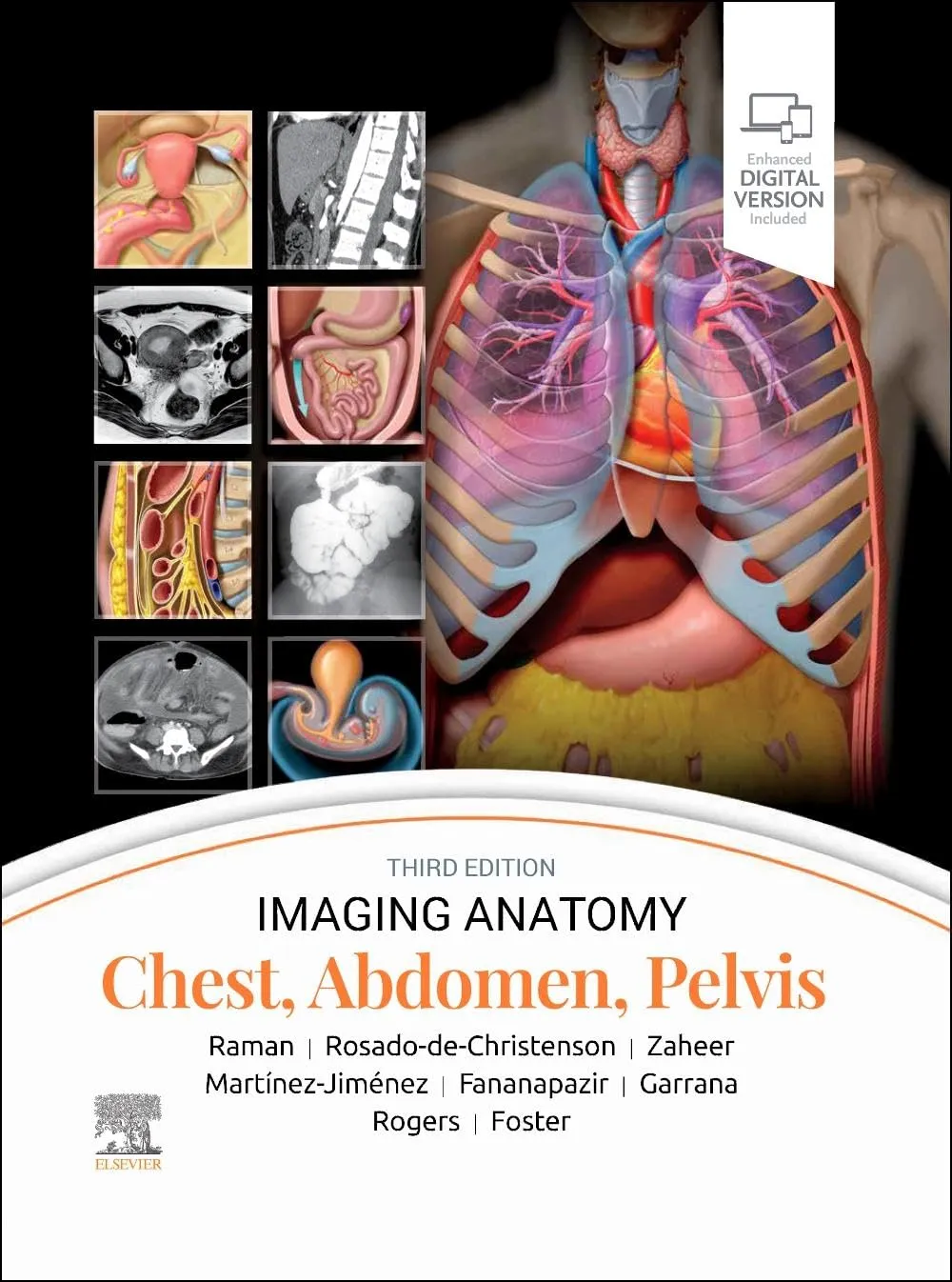
Imaging Anatomy: Chest, Abdomen, Pelvis-3E
by Raman
Highlights

9780443118005
ISBN
Raman
Author

1192
Pages

8000 gm
Weight

English
Language

2023
Year

N/A
Edition

Hardcover
Binding
₹25396
₹28218
This richly illustrated and superbly organized text/atlas is an excellent point-of-care resource for practitioners at all levels of experience and training. Written by global leaders in the field, Imaging Anatomy: Chest, Abdomen, Pelvis, third edition, contains specifics about radiographic, multiplanar, high-resolution, and cross-sectional body imaging along with thousands of relevant examples to give busy clinicians quick answers to imaging anatomy questions. This must-have reference employs a templated, highly formatted design; concise, bulleted text; and state-of-the-art images throughout that identify characteristic normal imaging findings and anatomic variants in each anatomic area, offering a unique opportunity to master the fundamentals of normal anatomy and accurately and efficiently recognize pathologic conditions. Contains nearly 2,800 print and online-only images, including all relevant imaging modalities, 3D reconstructions, and detailed, high-resolution medical drawings that together illustrate the fine points of imaging anatomy Reflects new understandings of anatomy due to ongoing anatomic research as well as new, advanced imaging techniques Offers new content on the anatomic basis for thoracic developmental abnormalities, anatomic variants of systemic and pulmonary vasculature, and the PI-RADS system and clinical implications of MR for prostate cancer Contains new and updated images of the chest wall musculature with CT and MR examples; abdominal imaging best practices, including the application of body MR in the abdomen and pelvis; and the different modalities used for GU/GYN imaging, specifically retrograde urethrography and MR for specific disease diagnosis Depicts common anatomic variants and covers the common pathological processes that manifest with alterations of normal anatomic landmarks Features representative pathologic examples to highlight the effect of disease on human anatomy Presents essential text in an easy-to-digest, bulleted format, enabling imaging specialists to find quick answers to anatomy questions encountered in daily practice Includes an eBook version that enables you to access all text, figures, and references with the ability to search, customize your content, make notes and highlights, and have content read aloud Although the anatomy of the chest, abdomen, and pelvis does not change, the 3rd edition of IA: CAP includes updates to each of the book's 3 anatomical sections, including: Text and imaging updates that tie common as well as very important but potentially uncommon current clinical issues to anatomy descriptions and examples as related to best practices for radiology reporting Updated images across all 3 sections of the book Anatomic basis for some thoracic developmental abnormalities Anatomic variants of systemic and pulmonary vasculature Updated drawings of the chest wall musculature with CT and MR examples Abdominal imaging best practice updates More emphasis on the roles of different modalities used for GU/GYN imaging, specifically retrograde urethrogram and MR for specific disease diagnosis Additional details added on the PIRADS system and clinical implications of MR for prostate cancer
Online store of medical books
Discover a comprehensive range of medical books at our online store. From anatomy and physiology to the latest clinical guidelines, we've got you covered.
Trusted by students, educators, and healthcare professionals worldwide. Browse top publishers and expert-authored titles in every medical specialty. Enjoy fast shipping, secure payments, and easy returns. Your one-stop destination for quality medical knowledge at your fingertips.
Whether you're preparing for exams or expanding your clinical expertise, our curated collection ensures you have the right resources at hand. Dive into detailed illustrations, case studies, and up-to-date research that enhance your understanding and practical skills.
We regularly update our inventory to include the latest editions and newly released titles, helping you stay current in the ever-evolving medical field. Our advanced search and filtering tools make finding the perfect book quick and hassle-free.
Join our community of lifelong learners and medical enthusiasts. Sign up for exclusive discounts, early access to new arrivals, and personalized book recommendations tailored to your professional interests.