
Home
|
Products
|
9789356962026
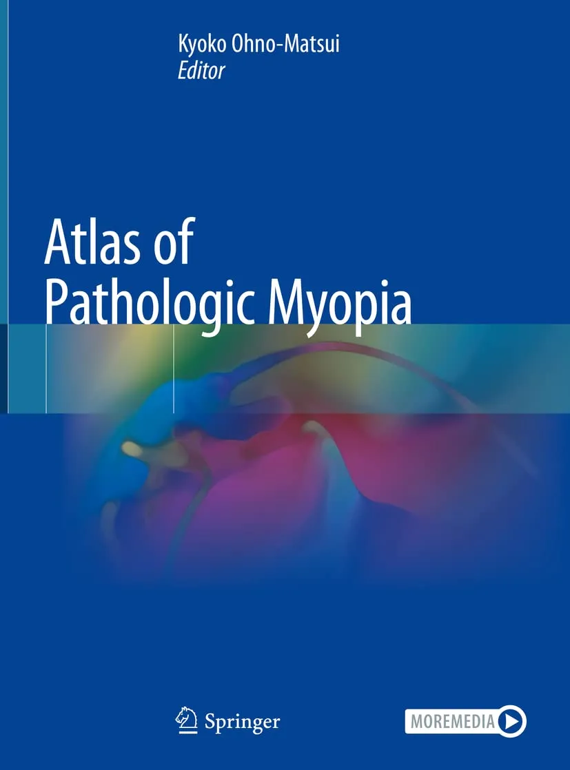
Atlas Of Pathologic Myopia | Hardcover
by Pjmp-Matsui K.
Highlights

9789811542602
ISBN
Pjmp-Matsui K.
Author

202
Pages

845 gm
Weight

English
Language

2020
Year

1st Edition
Edition

Hardcover
Binding
₹18188
₹20209
This Atlas provides many beautiful images obtained with state-of-the-art technologies, including optical coherence tomography (OCT), OCT angiography, fundus autofluorescence, and wide-field fundus imaging, as well as traditional images and fluorescein/ICG angiograms. Gathered at the worlds largest High Myopia Clinic, the images are based on the long-term follow-up data of more than 6,000 patients from Japan and abroad. Recent advances in imaging technologies have yielded many new observations and allowed us to detect new lesions, e.g. myopic traction maculopathy (or macular retinoschisis) and dome-shaped macula. An especially interesting aspect: the images obtained by ˜3D MRI of the eye and ˜ultra wide-field OCT to visualize staphylomas. These techniques were established by the editors group and make it possible to record the entire shapes of the eye, offering a scan width of up to 23 mm and scan depth of 5 mm. They have since been used to visualize posterior staphyloma, which was previously impossible to view because it spanned such a wide range of the eye. In addition, readers will learn what types of eye deformity occur in pathologic myopia and how they damage the macula/optic nerve. With this Atlas, readers will learn how to accurately diagnose each lesion of pathologic myopia, how eye deformity causes blinding complications, and how to identify patients with a poor prognosis. In short, it provides essential information that cant be found elsewhere.
Online store of medical books
Discover a comprehensive range of medical books at our online store. From anatomy and physiology to the latest clinical guidelines, we've got you covered.
Trusted by students, educators, and healthcare professionals worldwide. Browse top publishers and expert-authored titles in every medical specialty. Enjoy fast shipping, secure payments, and easy returns. Your one-stop destination for quality medical knowledge at your fingertips.
Whether you're preparing for exams or expanding your clinical expertise, our curated collection ensures you have the right resources at hand. Dive into detailed illustrations, case studies, and up-to-date research that enhance your understanding and practical skills.
We regularly update our inventory to include the latest editions and newly released titles, helping you stay current in the ever-evolving medical field. Our advanced search and filtering tools make finding the perfect book quick and hassle-free.
Join our community of lifelong learners and medical enthusiasts. Sign up for exclusive discounts, early access to new arrivals, and personalized book recommendations tailored to your professional interests.